This is an archival copy of the Visualization Group's web page 1998 to 2017. For current information, please vist our group's new web page.

| |
Early Abstract for IEEE Vis99 Submission
We describe a prototype system in which a virtual caliper is used to measure three dimensional relationships between objects perceived by an observer from stereo image pairs. The qualitative results from this experiment illustrate that stereo imaging combined with motion parallax allow a researcher to measure three dimensional relationships in data acquired from an electron microscope. Of particular concern are issues relating to consistency of imaging models between the virtual and real cameras. This system shows promise, but does raise many interesting questions.
Problem Definition
The basic thesis of this work is that the human perception system is capable of formulating a sufficiently meaningful impression of shape information from limited information for the purpose of measuring angles between perceived objects.
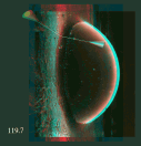
Images from "different viewpoints" are obtained from the instrument by tilting the specimen. These images can be viewed in a stereoscopic format, such as anaglyph or multibuffered, and observers have the impression of viewing a three dimensional object.
Similarly, a computer graphics based representation of a measuring device, such as a virtual caliper or protractor, can be rendered using virtual cameras positioned in such a way as to approximate the viewing geometry of a human observer with binocular vision. The final stereoscopic image of the virtual measuring device also appears to have the qualities of a three dimensional object.
Assuming that an observer can percieve three dimensional structure from stereo image pairs obtained from an instrument, and can perceive three dimensional structure from artifical computer graphics renderered from slighly different perspectives, the question that follows is ask if an observer can use a virtual device to measure relationships perceived in stereo image pairs.
A paper titled Measurement of Perceived Objects was accepted for publication and presentation in the Late Breaking Hot Topics section of IEEE Visualization 1999.
December 13, 1999
In the previous work, we focused on measurement of angles between perceived objects. In that study, a marker droplet was placed upon the surface of lung tissue, then imaged using a scanning electron microscope. The relative angle between the droplet and the lung tissue was believed to be indicative of the amount of surface tension on the lung tissue itself.
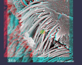
Based upon the success of the earlier project in which VR technology was combined with stereo microscopy, researchers at LBL would like to attempt to measure lengths of structures. The stereo image below shows a lung cell with cilia, fine hairlike structures whose function is to move debris along the surface of the lung while immersed in a bath of liquid. The image shows at least two different lengths of cilia, with glimpses of a very short, third layer.

|
|
Anaglyph stereogram of electron microscope images. Using red-blue glasses, place the red lens over the left eye. |
Preliminary investigations in our visualization laboratory lead us to believe that it will be possible to measure lengths of some of the cilia. Our approach will be to use a spline-based curve editing system, where the spline can be manipulated by a user in 3D, the geometry (the spline itself) is rendered in 3D stereo simultaneous with presentation of stereo images obtained from the microscope.